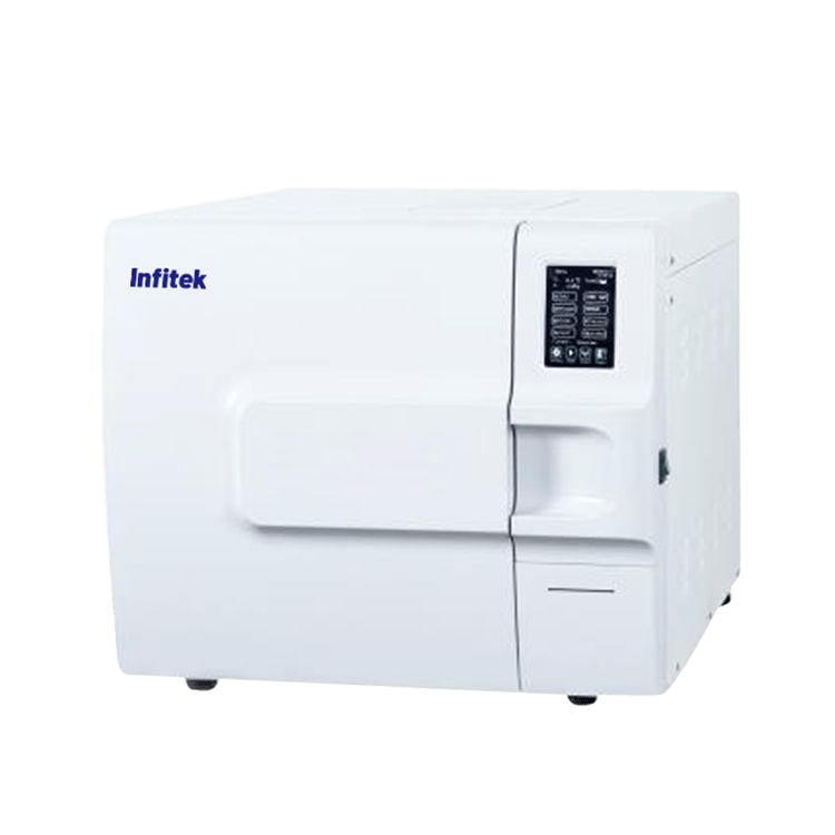
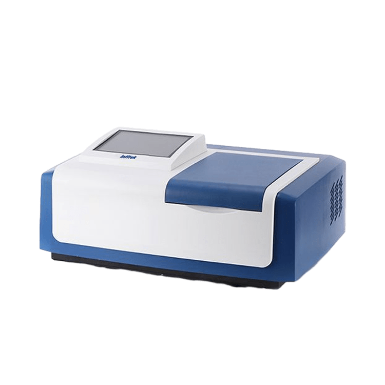
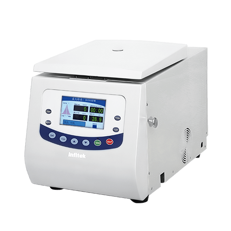
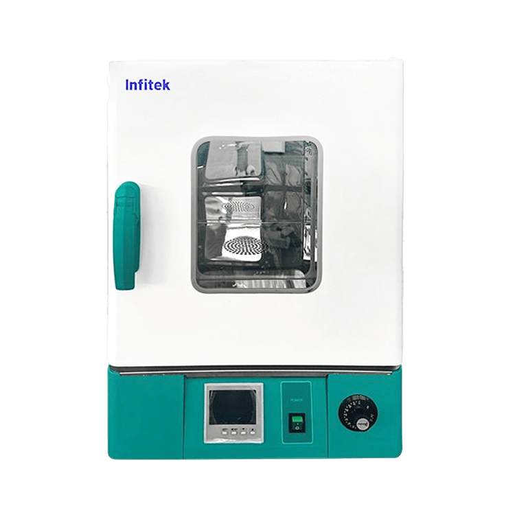
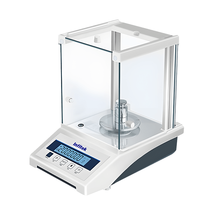
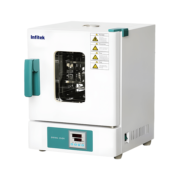
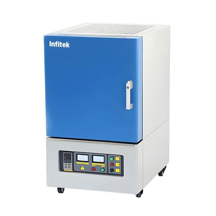
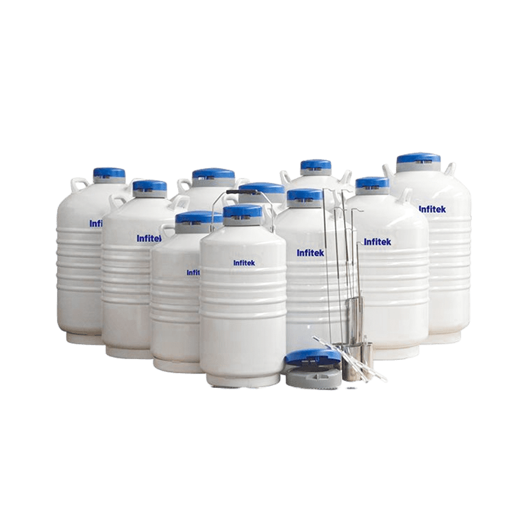
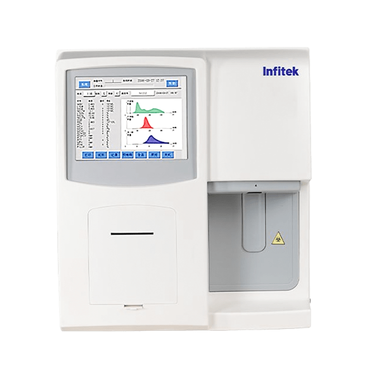
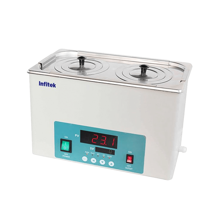
Hot Products

About Infitek
Founded in 2010, with the mission of continuously improving the intelligence, precision, safety and convenience of the laboratory, Infitek is a professional manufacturer driven by innovation and service in laboratory and Medical field, our company is certified by ISO9001, ISO13485 and Intellectual Property Management System.
News
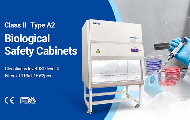
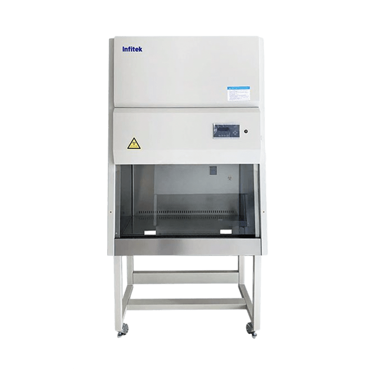
Enhancing Laboratory Safety with Biosafety Cabinets
In the realm of scientific research and experimentation, safety is paramount. Ensuring the [...]
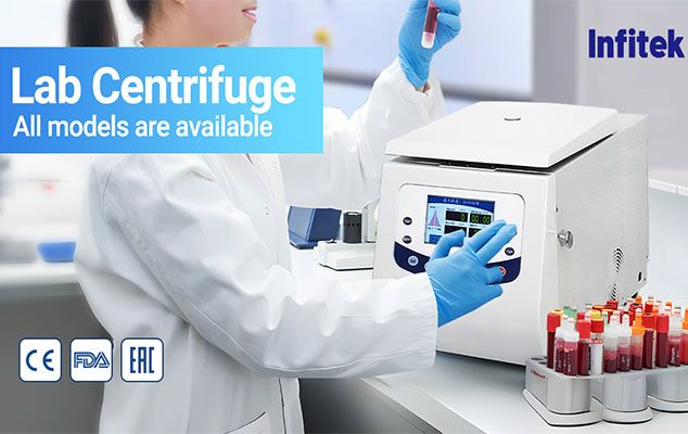
A Comprehensive Guide to Choosing the Right Laboratory Centrifuge
Introduction: Selecting the appropriate laboratory centrifuge is a crucial decision that can significantly [...]
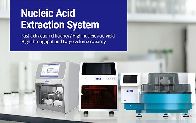
Streamlining Nucleic Acid Extraction in the Lab: The Role of Automated Extractors
In the dynamic landscape of molecular biology and diagnostics, efficient nucleic acid extraction [...]
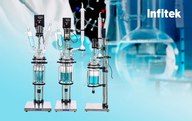
The Vital Role of Glass Reactors in Laboratory Research
In the realm of chemical synthesis and experimentation, the glass reactor stands [...]
Get Social