
- High resolution medical 15.6-inch display
- Spectrum envelope function
- One-click automatic optimization
- Rich user-defined functions
- Built-in large capacity lithiumbattery (detachable)
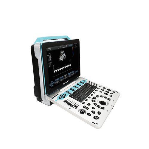
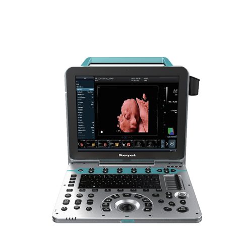
Color Doppler Ultrasound Scanner, Laptop, US-HCL5
Description
It is equipped with a high-resolution medical display, adopts multi-beam parallel technology and sub-array element transducers, and its superior image clarity perfectly meets the needs of women’s health care.
Features
- Brilliant ergonomics
High resolution LED with tilting funcitonality
User friendly keyboard and controls
- Excellent image quality
Tissue Harmonic Imaging(THI)enhancing contrast resolution
Quick image optimization by IP(Image Processing)
- segment TGC allowing delicate image adjustment
- Powerful workflow
One-key save, user-defined keys bring ng great efficiency to your daily diagnosis.
- Clinical Versatility
6 probes with abundant measurement packages covering traditional ultrasound applications and emerging fields such as urology, MSK and anesthesia.
Specifications
| Model | US-HCL5 |
| Main Parameters | |
| Ultrasonic Host Operating System | Windows 8 operating system |
| Description | Spectrum pulse Doppler |
| Direction energy Doppler | |
| Real time three synchronization | |
| Space composite imaging | |
| Tissue harmonic imaging | |
| 2B / 4B imaging mode | |
| Language | English/Russian/Spanish/French |
| Display | 15 inches, high definition LED |
| 0-180°Angle adjustable | |
| Physical Clipboard | Save the image on the left side of the screen, which can be directly saved or deleted. |
| Function | On-the-spot upgrade function |
| Trapezoidal imaging function | |
| Presupposition | For different inspection of the viscera, preset the inspection conditions for the best image, reduce the adjustment of the operation, and the commonly used external adjustment and combination regulation. |
| Signal Probe Interface | With |
| Probes | |
| Convex Probe | 2.5MHz/3.0MHz/3.5MHz/4.0MHz/H4.0MHz/H5.0MHz,(Depth: 20-317MM) |
| Linear Probe | 6.0MHz/7.5MHz/8.5MHz/10.0MHz/H10.0MHz,(Depth:20-110MM) |
| Transvaginal probe
|
4.5MHz/6.0MHz/7.0MHz/9.0MHz/H8.0MHz, (Depth:30-111MM) |
| Phased Array Probe | 2.5MHz/3.0MHz/3.5MHz/4.0MHz/H3.0MHz/H4.0MHz,(Depth:30-371MM) |
| Micro-Convex Probe(R11) | 4.5MHz/6.0MHz/7.0MHz/9.0MHz/H8.0MHz,(Depth:30-111MM) |
| Micro-Convex Probe(R20) | 2.5MHz/3.0MHz/3.5MHz/4.0MHz/H4.0MHz/H5.0MHz,(Depth:30-23.7MM) |
| The above probes have harmonic frequencies.
Each probe has a selection of specialist and organ modes for quick access for diagnostic. |
|
| Two-Dimensional Imaging Mode | |
| Gain | 0-100, step 1 visible adjustable |
| TGC | 8 segment adjustable |
| Dynamic Range | 20-280dB 20 level visual adjustable |
| Pseudo Color | 12, visible and adjustable |
| Sound Power | 5% to 100%, step 5%, visible and adjustable |
| Body Mark | ≥6 kinds |
| Max.Focus Number: | ≥6. adjustable |
| Gray Scale | 0-7 level visible visible and adjustable |
| Filtering | ≥5 kinds |
| Scanning Range | 50%-100% |
| Frame Correlation | 0-4 level, visible and adjustable |
| Display Content | The screen shows 14 forms of real time display of power, probe frequency, dynamic range, pseudo color, grayscale and so on. |
| Color Imaging Mode | |
| Colour Deflection | Workable |
| Color Frame Correlation | 12 levels, visible and adjustable |
| Color Map | 7, visible and adjustable |
| Color Reversal | Adjustable |
| B/C Split Screen Synchronous Display Function | Workable |
| Color Baseline | 7, visible and adjustable |
| Color Line Density | Adjustable |
| Spectrum Doppler Mode | |
| Sampling Volume Angle Correction | -80°~80° adjustable |
| Sampling Volume | 0.5mm-20mm visible and adjustable |
| Frequency | ≥5, visible and adjustable |
| Baseline | 8 steps adjustable |
| Smooth | 8 steps adjustable |
| Display Layout | ≥4 kinds, visible and adjustable |
| Pseudo Color | ≥ 7, visible and adjustable |
| Speed Scale | 3-2288cm/s |
| Spectrum Envelope Function | Real time automatic spectrum envelope, manual spectrum envelope, and so on. |
| System Automatic Analysis and Display | PSV, EDV, RI, PI, S/D, ACC, HR and other kinds of data |
| Measurement and Analysis Function | |
| Measurement | General measurement distance, area, angle, time, slope, heart rate, velocity, acceleration, neck hyaline layer, spectrum tracing, resistance index / pulsatility index, etc. |
| Obstetric Measurement | Weight measurement formula ≥13 options |
| Measuring Line | The color and line type of the measuring line adjustable
(including activating the color and completing the color). |
| Location and Font Size | The measurement results show that the location and font size adjustable.
|
| Professional Software Package | Abdomen, obstetrics and Gynecology, small organs, carotid, Urology, orthopedics, peripheral blood vessels, heart. |
| Graphic and Text Management System | |
| Image Storage Format | BMP DICOM JPEG DEFAUIT |
| Host | It built in ≥128G solid state hard disk to start fast and stable |
| Movie Playback | ≥ 600 frames |
| Internal Patient File Information Management System | Record patient number, name, check number, check date and so on, and can be searched and managed by numbering, checking number, name and so on. |
| Report | Type of report≥ 18 |
| One key fast report graphic and text management | |
| Interface | |
| USB Port | 2 |
| Audio Port | 1 |
| LAN Port | 2 |
| HDMI Port | 1 |
| Configuration | |
| Main Unit | 1pc. |
| Probes | Convex probe(Standard), Linear probe(Optional), Micro-convex probe of R20(Optional), Phased array probe(Optional), Trans-vaginal probe(Optional), Volume probe(Optional) |
| Thermal Video Printer | Optional |
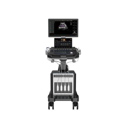
| Mobile Trolley | Optional |
| Dimension&Weight | |
| External Dimension | 375*350*480mm |
| N.W./G.W. | 8.6kg/13.12kg |
| Shipping Dimension | 560*390*570mm |


Get Social