- Ergonomic design
- Windows embedded ultrasonic running platform
- DICOM function, can be directly connected with the hospital PACS system
- Automatic spectrum measurement
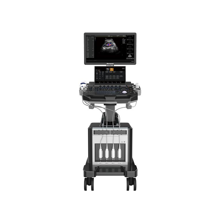
Trolley Color Doppler Ultrasound Machine, US-C30
Description
The US-C30 Trolley Color Doppler Ultrasound Machine is equipped with a high-resolution medical display, using multi-beam parallel technology and sub-array element transducers. Its excellent image clarity and rich measurement package perfectly meet the needs of women’s health care.
Features
- Expandable features
Four fully activated probe interfaces. USB image storage export. Can connect external monitor and printer.
- High efficiency
Built-in file information management system can record the number, name, check number, check date, and so on, and can search through the number, check number, name,etc.
The diagnostic report can be edited, ultrasound diagnostic image can be embedded in the report, and directly printed.
With DICOM function, can be directly connected with the hospital PACS system.
- Simpleoperation
Integral small keyboard, easy to operate. Probe storage tank free combination.
Automatic spectrum measurement.
The obstetric model can modify a variety of measurement combinations
- Humanized design
Main screen is 21.5-inch and auxiliary screen is 13.3-inch medical ultrasonic touchscreen , to meet the needs of doctors from different angles.
The full use of touch screen can reduce inaccurate operation caused by button contact and pressing, and minimize user fatigue.
Windows embedded ultrasonic running platform.
Specifications
| 1. Product Name: US-C30 Trolley Color Doppler Ultrasound System |
| 1. 1 Structure type: Dual-screen trolley type |
| 2. Purpose and Applications
2. 1 To meet the basic hospitals in the abdomen, obstetrics, gynecology, heart, urinary system, small organs, superficial, blood vessels, pediatrics, neonatal, muscle, physical examination and other inspections and diagnosis. |
| 3.Main Specifications and System Overview |
| 3. 1 Operating system: Windows 10
3.2 Pulse Wave Doppler 3.3 Directional Energy Doppler 3.4 Real-time triple synchronization 3.5 Spatial Composite Imaging 3.6 Tissue Harmonic Imaging 3.7 2B/4B Imaging 3.8 Support system languages: English, French, Russian, Spanish. 3.9 Standard monitor size: 21.5-inch, high-definition medical grade LCD display, and 13.3inch touch-screen. 3. 10 Integrated clipboard: display the saved images at the bottom of the screen, which can be directly transferred or deleted 3. 11 With on-site upgradeable functions 3. 12 Preset conditions: For different inspections, preset inspection conditions for optimized images, reducing adjustments during operation, and commonly required external adjustments and combined adjustments 3. 13 Support real-time 3D imaging function (3D/4D/5D) (optional) 3. 14 Probe ports: 4 active 3. 15 Trapezoidal Imaging 3. 16 One-key auto optimization |
| 4. Multiple Frequency of Different Probes |
| Convex probe: 2.0MHz/2.5MHz/3.0MHz/3.5MHz/4.0MHz/5.5MHz/H4.0MHz/H5.0MHz,(depth 30-255mm)
Linear probe: 6.0MHz/7.5MHz/8.5MHz/ 10.0MHz/ 12.0MHz/H10.0MHz, (depth 20-128mm) Trans-vaginal probe: 4.5MHz/5.0MHz/6.0MHz/6.5MHz/7.0MHz/9.0MHz/H8.0MHz, (depth 30- 156mm) Phased array probe: 2.0MHz/2.5MHz/3.0MHz/3.5MHz/4.0MHz/5.0MHz/H3.0MHz/H4.0MHz, (depth 100-244mm) 4D Volume probe: 2.0MHz/3.0MHz/4.0MHz/4.5MHz/5.5MHz/6.0MHz/H5.0MHz, (depth 30-237mm) (optional) Micro convex probe (R15): 4.0MHz/6.0MHz/7.0MHz/8.0MHz/H8.0MHz, (depth 30- 111mm) Micro convex probe (R20) :4.5MHz/6.0MHz/7.0MHz/9.0MHz/H8.0MHz,(depth 30- 111MM) Rectal probe:4.0MHz/6.5MHz/9.0MHz/H8.0MHz ,(depth 20-111MM) |
| 5. 2D Mode |
| 5. 1 Gain: 0-100, single step 1, visible and adjustable
5.2 TGC: 8 segment adjustable 5.3 Dynamic range: 20-280dB, 20 levels visible and adjustable 5.4 Pseudo color: 12 kinds, visible and adjustable 5.5 Sound power: 5%-100%, single step 5%, visible and adjustable 5.6 Body mark: 6 kinds 5.7 Number of focus: max. 6 5.8 Gray Scale: 0-7, visible and adjustable 5.9 Filtering: 0-4 5. 10 Scanning range: 50%- 100% 5. 11 Frame related: 0-4, visible and adjustable 5. 12 Real-time display of 14 parameters such as sound power, probe frequency, dynamic range, pseudo color, gray scale, etc., which are adjustable 5. 13 Scanning line density: high, medium and low adjustable 5. 14 Noise reduction: 0- 14 5. 15 Pseudo-color spectrum: 0- 11 5. 16 Support frequency compound |
| 6. Color Imaging Mode |
| 6. 1 Color deflection: available
6.2 Color frame related: 0- 12 levels, visible and adjustable 6.3 Color map: 0-3 levels, visible and adjustable 6.4 Color flip: adjustable 6.5 B/C split screen synchronous display function: available 6.6 Color baseline: 11 levels, visible and adjustable 6.7 Color line density: high and low adjustable 6.8 Wall filter: 0-5 level adjustable |
| 7. Pulse Wave Doppler Mode |
| 7. 1 Sampling volume angle correction: -80°~80° adjustable
7.2 Sampling volume: 0.5mm-20mm visible and adjustable 7.3 Frequency: 2.5MHz and 3.0MHz visible and adjustable 7.4 Baseline: 11 levels adjustable 7.5 Pseudo-color spectrum: 0-5 7.6 Display layout: 4 kinds, visible and adjustable 7.7 Pseudo-color: 6 kinds adjustable *7.8 Speed scale: 3-2288cm/s 7.9 Spectrum envelope function: real-time automatic spectrum envelope, manual spectrum envelope and other modes are available, the system automatically analyzes and displays: PSV, EDV, RI, PI, S/D, ACC, HR and other data. 7. 10 Grayscale: 0-7 7. 11 Wall filter: 0- 12 7. 12 Dynamic range: 10-95dB, single step 5 7. 13 Noise reduction: 0-60 7.14 Volume: 0-100 |
| 8. Real Time 3D Imaging Mode |
| 8. 1 Fast angle: support 0°, 90°, 180°, 270° rotation of the 3D window image
8.2 Display layout: support “double frame”, “four frame”, “single frame” image display 8.3 Reconstruction modes: RealSkin, surface, Max, Min, XRax five reconstruction modes 8.4 Pseudo-color display: support 0-7 level adjustment 8.5 Image magnification: support 5 levels 8.6 Contrast: 0%- 100% 8.7 Threshold: 0%- 100% 8.8 Smooth: 3 levels adjustable 8.9 Pseudo-color: 7 levels adjustable 8. 10 X-axis, Y-axis, and Z-axis rotation support adjustable 8. 11 Brightness: 0%- 100% |
| 9. 4D Imaging Mode(optional) |
| 9. 1 Fast angle: support 0°, 90°, 180°, 270° rotation of the 3D window image
9.2 Display layout: support “double frame”, “four frame”, “single frame” image display 9.3 Reconstruction modes: RealSkin, surface, Max, Min, XRax five reconstruction modes 9.4 Pseudo-color display: 0-7 levels adjustable 9.5 Image magnification: 5 levels 9.6 Contrast: 0%- 100% 9.7 Threshold: 0%- 100% 9.8 Smooth: 3 levels adjustable 9.9 Pseudo-color:7 levels adjustable 9. 10 X-axis, Y-axis, and Z-axis rotation adjustable 9. 11 Line density: 2 levels adjustable |
| 10. Measurement and Analysis Functions: |
| 10. 1 General measurement of distance, area, angle, time, slope, heart rate, speed, acceleration, neck transparent layer, spectrum trace, resistance index/pulsation index, etc.
10.2 Obstetric measurement: weight measurement formula: 13 kinds for option. 10.3 The color and type of the measuring line can be adjusted(including the activation color and the completion color). 10.4 The display position and font size of the measurement results can be adjusted. 10.5 Measurement packages: abdomen, obstetrics, urology, etc. |
| 11. Picture And Text Management System: Picture saving format: BMP/DICOM/JPEG as default |
| 11. 1 Built-in 128GB solid-state hard drive in the main unit to start fast and stable.
11.2 Cine loop: 600 frames 11.3 Built-in file information management system: with record number, name, inspection number, inspection date, etc., and can search and manage by number, inspection number, name, etc. 11.4 Report types: 17 kinds 11.5 One-key quick report graphic management |
| 12. Interfaces |
| 4*USB ports, 1*Video, 1*S-Video, 1*DVI, 1*HDMI, 1*RJ-45 |
| 13.Configurations: |
| 13.1 Trolley Color Doppler Ultrasound System Host*1set
13.2 Probe options: Convex probe linear probe Micro-convex probe R20 Phased array probe Trans-vaginal probe 4D volume probe (optional) 13.3 Optional accessories: Thermal printer, puncture guide frame, gel warmer etc. |


Get Social